Spinal L1 Lumbar vertebrae l1 rib human anatomy preview illustration
If you are searching about Innervation of the lumbar spine. (a) Diagram shows the innervation of you've came to the right page. We have 35 Pics about Innervation of the lumbar spine. (a) Diagram shows the innervation of like Lumbar Spine Injury L1-L5 | Spinal Cord, FE model of the lumbar spine. a (1) Intact model (L1-S1). Frontal and and also Anatomy of the lumbar spine | Healing touch | Pinterest | The o'jays. Read more:
Innervation Of The Lumbar Spine. (a) Diagram Shows The Innervation Of
 www.researchgate.net
 lumbar innervation facet joints nerve s1 vertebrae l5 l3 l4 nerves spinal t12 transverse medial dorsal inferior sacrum articular innervates
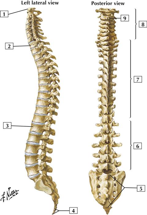
Whole Spine? - Denison Chiropractic
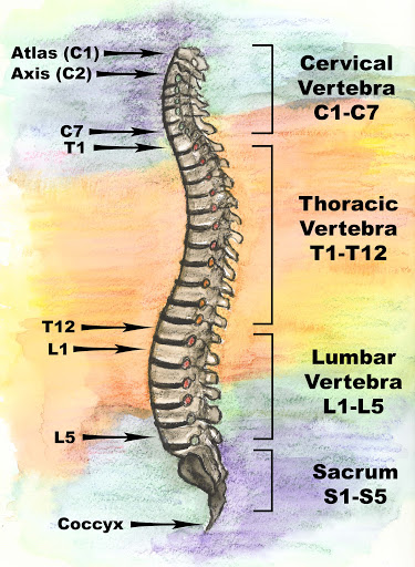 denisonchiropractic.com
 spine spinal c7 regions cervical bones diagram c4 c5 c3 c1 c6 l1 anatomy c2 l3 l2 cord l4 l5
Adjacent Segment Degeneration L1-L4 - TrialExhibits Inc.
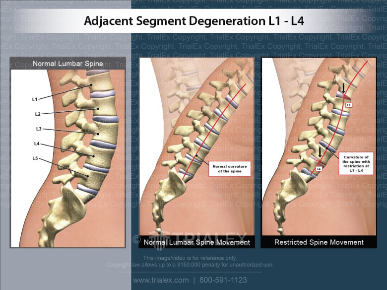 www.trialexhibitsinc.com
 l4 degeneration adjacent lumbar spinal
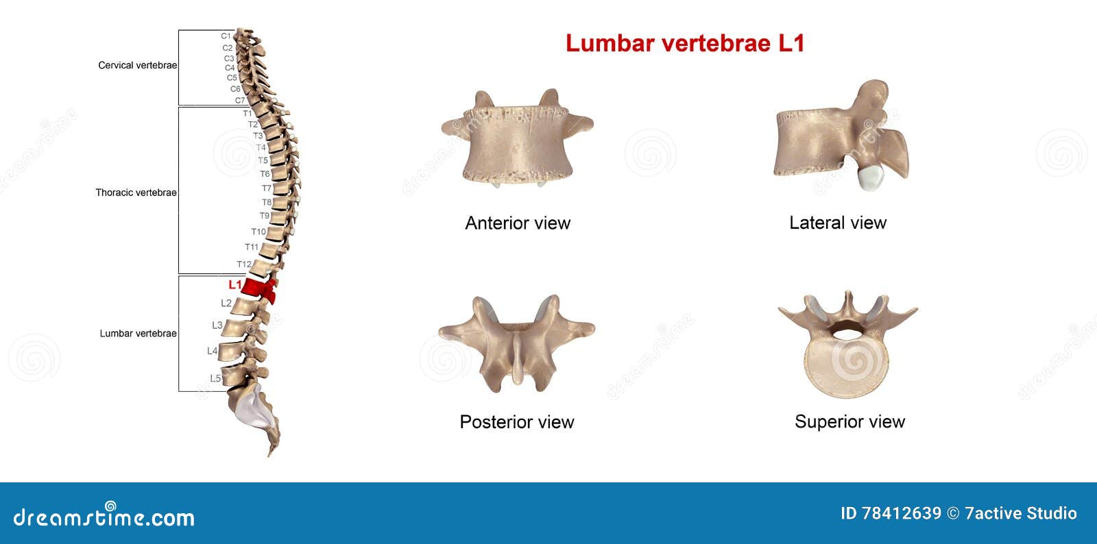
Lumbar Vertebrae L1 Stock Illustration. Illustration Of Bones - 78412678
 www.dreamstime.com
 lumbar l1 vertebrae vertebra human anatomy preview illustration similar
Spine Surgery - L5-S1 Spondylolisthesis With Spinal Cord Stock Photo
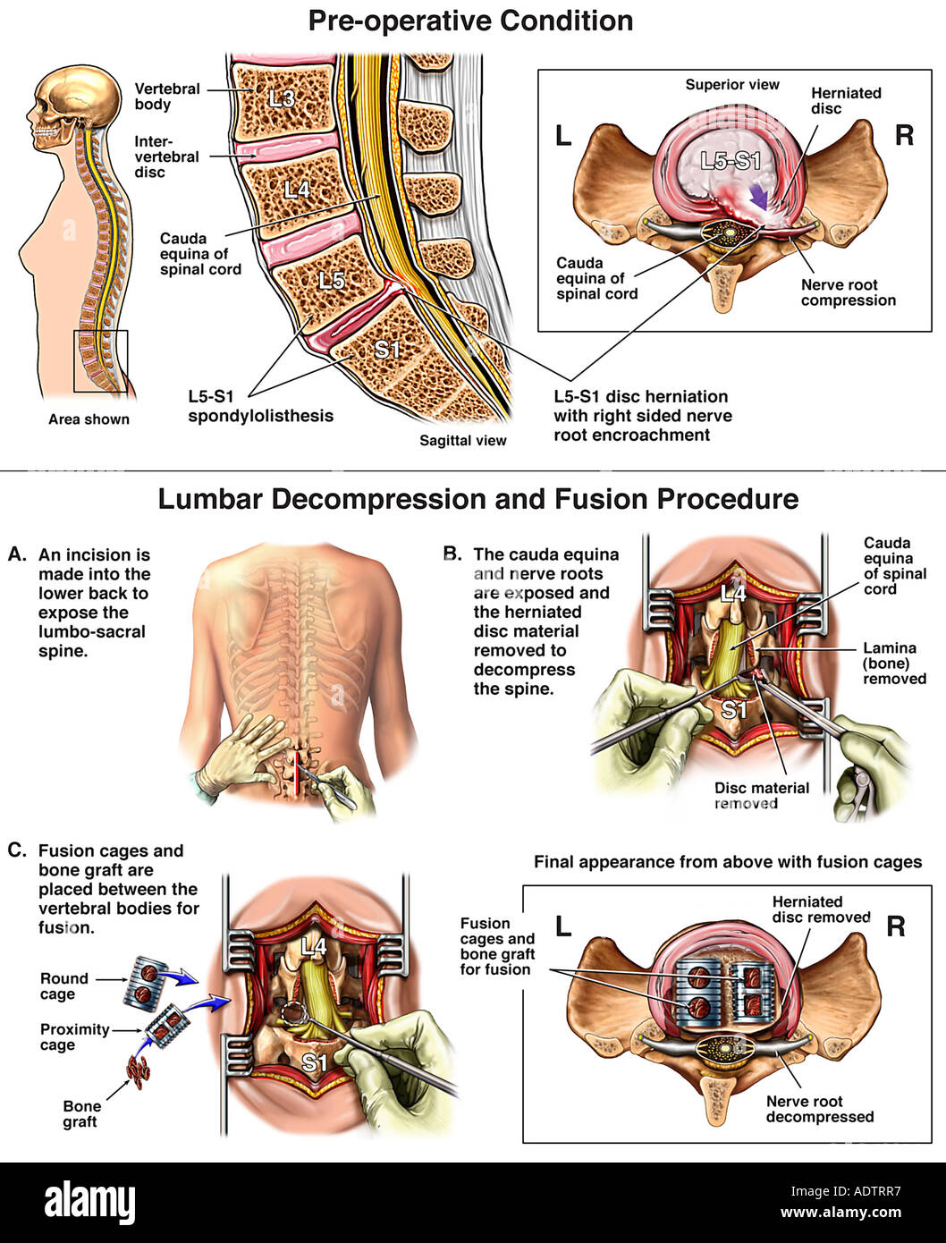 www.alamy.com
 l5 s1 surgery spinal spondylolisthesis cord decompression spine alamy
Anatomy Of The Lumbar Spine | Healing Touch | Pinterest | The O'jays
 www.pinterest.com
 lumbar spinal
AHP Suffolk > Home > Clinical Education > Spinal > Mechanical LBP
 ahpsuffolk.co.uk
 l1 spinal annulus fibrosus nuc nucleus ligament pulposus posterior longitudinal mechanical pll flava supraspinal isl ligamenta lbp
3D Model Of Spinal Segment L1-S1. ANN - Annulus Fibrosus, NUC - Nucleus
 www.researchgate.net
 spinal annulus nucleus fibrosus nuc pulposus ligament flava ligamenta posterior
L1 Vertebrae - Draw-shenanigan
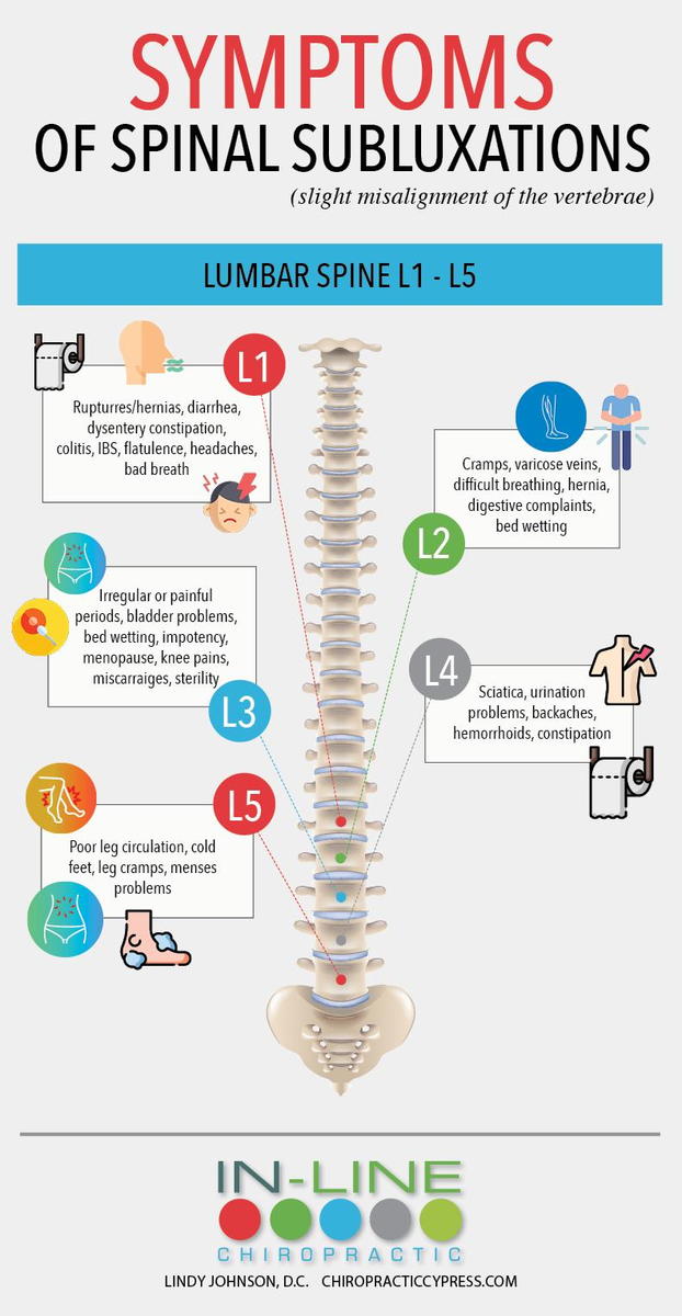 draw-shenanigan.blogspot.com
 l1 vertebrae symptoms misalignment spinal
L1 L2 Lumbar Spine Injuries | High Impact® Visual Litigation Strategies™
 www.highimpact.com
 l2 l1 spine lumbar injuries
Pin On Back Injuries & Spine Disorders | What's Causing My Back Pain
 www.pinterest.com
 fracture t12 compression vertebral thoracic spinal spine vertebrae vertebra lumbar fractures traumatologia ortopedia transitional nerve diagnosed mean
Lumbar Spine L1 Compression Fracture Trial Exhibit – Stock Trial Exhibits
 stocktrialexhibits.com
 fracture lumbar vertebra vertebral exhibits axial
30 Best Spinal L1 To L5 Images On Pinterest
 www.pinterest.com
 spinal nerve t12 l5
Lumbar Vertebrae L1 Stock Illustration. Illustration Of Bony - 81729174
 www.dreamstime.com
 lumbar vertebrae l1 rib human anatomy preview illustration
Lumbar Spine Injury L1-L5 | Spinal Cord
 www.spinalcord.com
 L1 Vertebrae - Draw-shenanigan
 draw-shenanigan.blogspot.com
 30 Best Images About Spinal L1 To L5 On Pinterest
 www.pinterest.com
 anatomy l1 innervation doctorlib info l5 colon medical atlas spinal anterior nerves human body
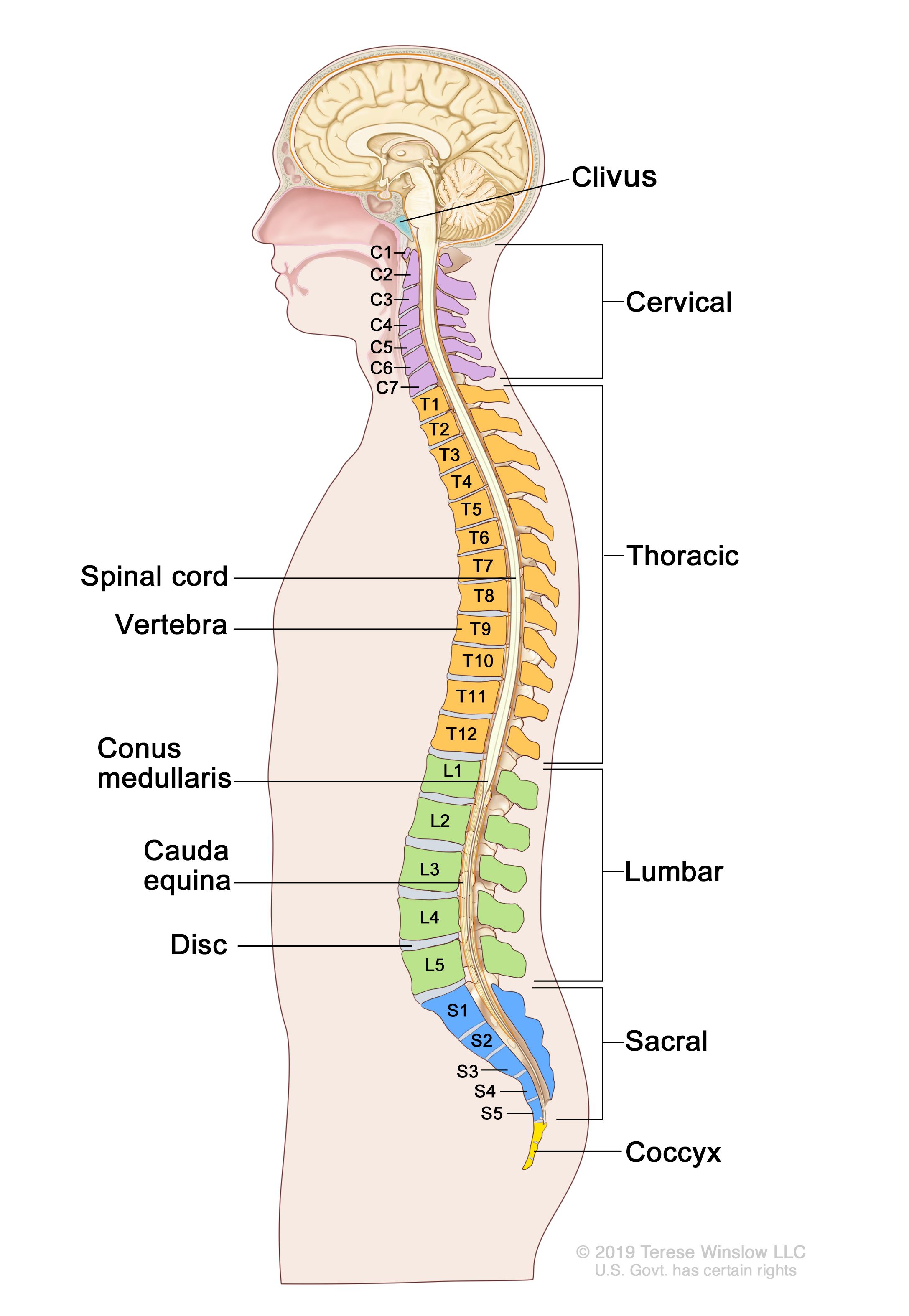
The Spinal Cord: The Spinal Cord Ends Between L1 And L2
 thespinalcordmaerio.blogspot.com
 spinal cord l2 l1 ends between
Back And Spinal Cord: Cards 2-1 To 2-21 | Basicmedical Key
 basicmedicalkey.com
 Lumbar Spinal Cord Injuries (L1-L5) Explained | SCI Progress
 sciprogress.com
 Lumbar Spine Injury L1-L5 | Spinal Cord
 www.spinalcord.com
 lumbar injury vertebrae spinal l5 vertebra numbness spinalcord
Exploration Of The Human Spinal Cord
 www.microscopy-uk.org.uk
 Anterior Wedge Fracture To The Vertebra | Rehab My Patient
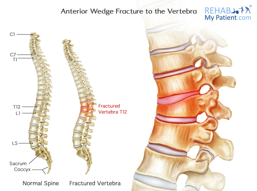 www.rehabmypatient.com
 wedge fracture anterior vertebra spine thoracic vertebrae lumbar fractures spinal link
Lumbar Vertebrae L1 Stock Illustration. Illustration Of Neck - 78412639
 www.dreamstime.com
 Lumbar Vertebrae: L1-5 Assembled
 www.netterimages.com
 lumbar l1 vertebrae spine netter anatomy labeled assembled pricing low other netterimages
Lumbar Spine Compression Fracture | RADIOLOGYPICS.COM
 radiologypics.com
 fracture spine columna l1 vertebral deformity radiologypics radiology radiograph xray posterior thoracalis frakturer lumbalis buyxraysonline radiography degenerative 보드 선택
PPT - Chapter 13 Spinal Cord, Nerves And Reflexes PowerPoint
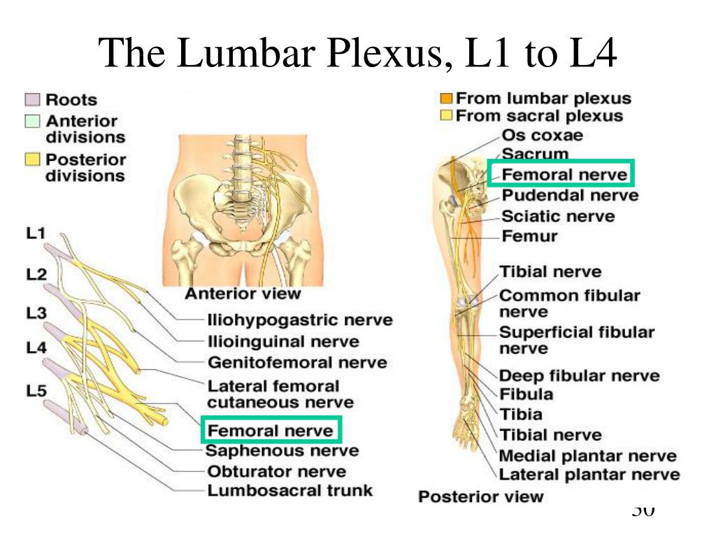 www.slideserve.com
 lumbar spinal plexus l1 l4 nerve nerves cord reflexes chapter injury
Illustration Of The Spinopelvic Parameters: T4–T12 Kyphosis, L1–L5
 www.researchgate.net
 t12 l1 l5 spinopelvic spinal kyphosis parameters lordosis pelvic lumbar thoracic inclination incidence
Lumbar Spine Injury L1-L5 | Spinal Cord
 www.spinalcord.com
 lumbar spinal l5 spine injury incomplete vertebrae spinalcord
FE Model Of The Lumbar Spine. A (1) Intact Model (L1-S1). Frontal And
 www.researchgate.net
 l1 spine frontal intact vertebrae l5 ligaments amaya palomar ligament
Injury Report: Lower Back Pain - Beautiful To The Core
 beautifultothecore.com
 spine lower pain lumbar surgery l1 vertebrae anatomy injury report vertebra successful bones beautifultothecore
30 Best Spinal L1 To L5 Images On Pinterest
 www.pinterest.com
 l5 l1 anatomy spinal medical pain gross doctorlib info
Figure 1 From Comparison Of 3D Spinal Motions During Stair-climbing
 www.semanticscholar.org
 spinal motions stair
TranS1 On Twitter: "All About The L5-S1 Lumbosacral Joint - Https://t
 twitter.com
 lumbar l5 s1 l4 trans1 lumbosacral spinal orthopedic verdite surgeon losangeles spinesurgeon
Spinal Cord Injury And How It Affects People | Back Up
 www.backuptrust.org.uk
 spinal vertebrae levels cervical lumbar thoracic injuries firstaidforfree backuptrust lesions occur
Lumbar vertebrae l1 stock illustration. illustration of bones. L1 spinal annulus fibrosus nuc nucleus ligament pulposus posterior longitudinal mechanical pll flava supraspinal isl ligamenta lbp. The spinal cord: the spinal cord ends between l1 and l2
if you are looking "id":318,"keyword_id":10,"name":"Spinal L1","scraped_at":"2023-02-11 14:02:34","created_at":"2023-02-11T14:02:34.000000Z","updated_at":"2023-02-11T14:02:34.000000Z","images":[ Spinal Cord","thumbnail":"https:\/\/tse4.mm.bing.net\/th?id=OIP.o49yJyXa_IPqiEynjMc-8AHaHa&pid=15.1","size":"700 x 700 \u00b7 jpeg","desc":"lumbar injury vertebrae spinal l5 vertebra numbness spinalcord","filetype":"jpg","width":700,"height":700,"domain":"www.spinalcord.com","created_at":null,"updated_at":null,"id":11166,"keyword_id":318,"url":"https:\/\/www.researchgate.net\/profile\/Amaya-Perez-Del-Palomar\/publication\/333263595\/figure\/fig1\/AS:761302314139648@1558520024050\/FE-model-of-the-lumbar-spine-a-1-Intact-model-L1-S1-Frontal-and-lateral-view-of-the.png","link":"https:\/\/www.researchgate.net\/figure\/FE-model-of-the-lumbar-spine-a-1-Intact-model-L1-S1-Frontal-and-lateral-view-of-the_fig1_333263595","title":"FE model of the lumbar spine. a (1) Intact model (L1-S1). Frontal and","thumbnail":"https:\/\/tse1.mm.bing.net\/th?id=OIP.nnV51Vb14kM-zIAy-5pT9wHaHP&pid=15.1","size":"850 x 831 \u00b7 png","desc":"l1 spine frontal intact vertebrae l5 ligaments amaya palomar ligament","filetype":"png","width":850,"height":831,"domain":"www.researchgate.net","created_at":null,"updated_at":null,"id":11167,"keyword_id":318,"url":"https:\/\/cdn.trialexhibitsinc.com\/library_media\/production\/d397c2b2be2178fe6247bd50fc97cff2\/c\/SP_C_0084-library_medium.jpg","link":"https:\/\/www.trialexhibitsinc.com\/library-item\/adjacent-segment-degeneration-l1-l4-1","title":"Adjacent Segment Degeneration L1-L4 - TrialExhibits Inc.","thumbnail":"https:\/\/tse4.mm.bing.net\/th?id=OIP.GZyQ4iJUr0lphLpzzoYZhwHaFi&pid=15.1","size":"780 x 584 \u00b7 jpeg","desc":"l4 degeneration adjacent lumbar spinal","filetype":"jpg","width":780,"height":584,"domain":"www.trialexhibitsinc.com","created_at":null,"updated_at":null, Spinal Cord","thumbnail":"https:\/\/tse2.mm.bing.net\/th?id=OIP.E9E68IpeSg-sJeVybviCUQHaDt&pid=15.1","size":"1200 x 600 \u00b7 jpeg","desc":"lumbar spinal l5 spine injury incomplete vertebrae spinalcord","filetype":"jpg","width":1200,"height":600,"domain":"www.spinalcord.com","created_at":null,"updated_at":null,"id":11169,"keyword_id":318,"url":"https:\/\/ai2-s2-public.s3.amazonaws.com\/figures\/2017-08-08\/002397aee7b393169c6d9b15d3769e47f3f3adbe\/3-Figure1-1.png","link":"https:\/\/www.semanticscholar.org\/paper\/Comparison-of-3D-spinal-motions-during-between-with-Lee-Desmoulin\/002397aee7b393169c6d9b15d3769e47f3f3adbe\/figure\/1","title":"Figure 1 from Comparison of 3D spinal motions during stair-climbing","thumbnail":"https:\/\/tse1.mm.bing.net\/th?id=OIP.qKlZRD-I74RtlYGIytJGFQHaGo&pid=15.1","size":"966 x 866 \u00b7 png","desc":"spinal motions stair","filetype":"png","width":966,"height":866,"domain":"www.semanticscholar.org","created_at":null,"updated_at":null,"id":11170,"keyword_id":318,"url":"https:\/\/pbs.twimg.com\/media\/CePAvWaW8AAqPEb.png:large","link":"https:\/\/twitter.com\/trans1\/status\/712625086193381377","title":"TranS1 on Twitter: \"All about the L5-S1 lumbosacral joint - https:\/\/t","thumbnail":"https:\/\/tse1.mm.bing.net\/th?id=OIP.XOFfX5jpKgx8afvt5rsQZgHaGy&pid=15.1","size":"1300 x 1193 \u00b7 png","desc":"lumbar l5 s1 l4 trans1 lumbosacral spinal orthopedic verdite surgeon losangeles spinesurgeon","filetype":"png","width":1300,"height":1193,"domain":"twitter.com","created_at":null,"updated_at":null,"id":11171,"keyword_id":318,"url":"https:\/\/ahpsuffolk.co.uk\/LinkClick.aspx?link=images%2fmlb1.jpg&tabid=320&portalid=1&mid=1290","link":"https:\/\/ahpsuffolk.co.uk\/Home\/ClinicalEducation\/Spinal\/MechanicalLBP.aspx","title":"AHP Suffolk > Home > Clinical Education > Spinal > Mechanical LBP","thumbnail":"https:\/\/tse1.mm.bing.net\/th?id=OIP.Mnf5Sy0pnqybqxfqEt7CiAHaFI&pid=15.1","size":"576 x 399 \u00b7 jpeg","desc":"l1 spinal annulus fibrosus nuc nucleus ligament pulposus posterior longitudinal mechanical pll flava supraspinal isl ligamenta lbp","filetype":"jpg","width":576,"height":399,"domain":"ahpsuffolk.co.uk","created_at":null,"updated_at":null,"id":11172,"keyword_id":318,"url":"https:\/\/nci-media.cancer.gov\/pdq\/media\/images\/797134.jpg","link":"https:\/\/draw-shenanigan.blogspot.com\/2021\/07\/l1-vertebrae.html","title":"L1 Vertebrae - Draw-shenanigan","thumbnail":"https:\/\/tse3.mm.bing.net\/th?id=OIP.glXuYvAP4jD50t6i_Ow14wHaKu&pid=15.1","size":"2175 x 3150 \u00b7 jpeg","desc":"","filetype":"jpg","width":2175,"height":3150,"domain":"draw-shenanigan.blogspot.com","created_at":null,"updated_at":null,"id":11173,"keyword_id":318,"url":"https:\/\/basicmedicalkey.com\/wp-content\/uploads\/2016\/06\/F000023u002-001-9780323185950.jpg","link":"https:\/\/basicmedicalkey.com\/back-and-spinal-cord-cards-2-1-to-2-21\/","title":"Back and Spinal Cord: Cards 2-1 to 2-21 ,"id":11174,"keyword_id":318,"url":"http:\/\/beautifultothecore.com\/wp-content\/uploads\/2015\/05\/spine.jpg","link":"http:\/\/beautifultothecore.com\/2015\/07\/11\/injury-report-lower-back-pain\/","title":"Injury Report: Lower Back Pain - Beautiful to the Core","thumbnail":"https:\/\/tse1.mm.bing.net\/th?id=OIP.gS3dZX1vEdkEz4_p-zMiAgAAAA&pid=15.1","size":"250 x 306 \u00b7 jpeg","desc":"spine lower pain lumbar surgery l1 vertebrae anatomy injury report vertebra successful bones beautifultothecore","filetype":"jpg","width":250,"height":306,"domain":"beautifultothecore.com","created_at":null,"updated_at":null,"id":11175,"keyword_id":318,"url":"https:\/\/image.slideserve.com\/735608\/the-lumbar-plexus-l1-to-l4-l.jpg","link":"https:\/\/www.slideserve.com\/carter\/chapter-13-spinal-cord-nerves-and-reflexes","title":"PPT - Chapter 13 Spinal Cord, Nerves and Reflexes PowerPoint","thumbnail":"https:\/\/tse1.mm.bing.net\/th?id=OIP.lpLM2zv1ECjCScEAEEP6RwHaFj&pid=15.1","size":"1024 x 768 \u00b7 jpeg","desc":"lumbar spinal plexus l1 l4 nerve nerves cord reflexes chapter injury","filetype":"jpg","width":1024,"height":768,"domain":"www.slideserve.com","created_at":null,"updated_at":null,"id":11176,"keyword_id":318,"url":"https:\/\/i.pinimg.com\/originals\/87\/e8\/89\/87e8899a68dcf7ebf928cf7ab9f56117.jpg","link":"https:\/\/www.pinterest.com\/pin\/119063983881518706\/","title":"Pin on Back Injuries & Spine Disorders ,"id":11177,"keyword_id":318,"url":"https:\/\/thumbs.dreamstime.com\/z\/lumbar-vertebrae-l-human-anatomy-five-rib-cage-pelvis-largest-segments-78412639.jpg","link":"https:\/\/www.dreamstime.com\/stock-illustration-lumbar-vertebrae-l-human-anatomy-five-rib-cage-pelvis-largest-segments-image78412639","title":"Lumbar vertebrae L1 stock illustration. Illustration of neck - 78412639","thumbnail":"https:\/\/tse1.mm.bing.net\/th?id=OIP.CD7A5xePTKowP746iGXf7AHaDx&pid=15.1","size":"1300 x 662 \u00b7 jpeg","desc":"","filetype":"jpg","width":1300,"height":662,"domain":"www.dreamstime.com","created_at":null,"updated_at":null,"id":11178,"keyword_id":318,"url":"https:\/\/denisonchiropractic.com\/wp-content\/uploads\/2020\/07\/full-spine.jpg","link":"https:\/\/denisonchiropractic.com\/2020\/07\/whole-spine\/","title":"Whole spine? - Denison Chiropractic","thumbnail":"https:\/\/tse4.mm.bing.net\/th?id=OIP.PYDHT4tuEt29Ho_txMu6ywAAAA&pid=15.1","size":"375 x 512 \u00b7 jpeg","desc":"spine spinal c7 regions cervical bones diagram c4 c5 c3 c1 c6 l1 anatomy c2 l3 l2 cord l4 l5","filetype":"jpg","width":375,"height":512,"domain":"denisonchiropractic.com","created_at":null,"updated_at":null,"id":11179,"keyword_id":318,"url":"https:\/\/www.backuptrust.org.uk\/wp-content\/uploads\/diagram-spine@3x.png","link":"https:\/\/www.backuptrust.org.uk\/spinal-cord-injury\/what-is-spinal-cord-injury","title":"Spinal cord injury and how it affects people ,"id":11180,"keyword_id":318,"url":"https:\/\/res.cloudinary.com\/high-impact\/image\/upload\/c_lpad,h_628,w_1200\/HI\/images\/projects\/13808-L1-L2-Lumbar-Spine-Injuries.jpg","link":"https:\/\/www.highimpact.com\/exhibits\/l1-l2-lumbar-spine-injuries","title":"L1 L2 Lumbar Spine Injuries ,"id":11181,"keyword_id":318,"url":"https:\/\/thumbs.dreamstime.com\/b\/lumbar-vertebrae-l-human-anatomy-five-rib-cage-pelvis-largest-segments-78412678.jpg","link":"https:\/\/www.dreamstime.com\/stock-illustration-lumbar-vertebrae-l-human-anatomy-five-rib-cage-pelvis-largest-segments-image78412678","title":"Lumbar vertebrae L1 stock illustration. Illustration of bones - 78412678","thumbnail":"https:\/\/tse1.mm.bing.net\/th?id=OIP.CEjZ3icXumEHiFZEsDsXHAHaDk&pid=15.1","size":"1600 x 771 \u00b7 jpeg","desc":"lumbar l1 vertebrae vertebra human anatomy preview illustration similar","filetype":"jpg","width":1600,"height":771,"domain":"www.dreamstime.com","created_at":null,"updated_at":null,"id":11182,"keyword_id":318,"url":"https:\/\/2.bp.blogspot.com\/-BiOxSZk9kjI\/T1bfMagNiUI\/AAAAAAAAAaI\/cuM0tVeJIiE\/s1600\/lumbarsagittal.gif","link":"https:\/\/thespinalcordmaerio.blogspot.com\/2015\/07\/the-spinal-cord-ends-between-l1-and-l2.html","title":"The Spinal Cord: The Spinal Cord Ends Between L1 And L2","thumbnail":"https:\/\/tse4.mm.bing.net\/th?id=OIP.kXT_OC1XV4xYD0A_-75bNAHaOv&pid=15.1","size":"200 x 398 \u00b7 gif","desc":"spinal cord l2 l1 ends between","filetype":"gif","width":200,"height":398,"domain":"thespinalcordmaerio.blogspot.com","created_at":null,"updated_at":null,"id":11183,"keyword_id":318,"url":"https:\/\/s-media-cache-ak0.pinimg.com\/736x\/02\/f2\/7c\/02f27ce5deaf7d0876e3034e93141ab9--spinal-nerve-spinal-cord.jpg","link":"https:\/\/www.pinterest.com\/kumarlimaye\/spinal-l1-to-l5\/","title":"30 best Spinal L1 to L5 images on Pinterest","thumbnail":"https:\/\/tse4.mm.bing.net\/th?id=OIP.iMlmP17vzVZYW1dpI5G8HwAAAA&pid=15.1","size":"198 x 396 \u00b7 jpeg","desc":"spinal nerve t12 l5","filetype":"jpg","width":198,"height":396,"domain":"www.pinterest.com","created_at":null,"updated_at":null,"id":11184,"keyword_id":318,"url":"http:\/\/cdn.shopify.com\/s\/files\/1\/0278\/6291\/products\/LumbarSpine_L1CompressionFracture_trialexhibit_1024x1024.jpg?v=1392773926","link":"http:\/\/stocktrialexhibits.com\/collections\/stock-trial-exhibits\/products\/lumbar-spine-l1-compression-fracture-trial-exhibit","title":"Lumbar Spine L1 Compression Fracture Trial Exhibit \u2013 Stock Trial Exhibits","thumbnail":"https:\/\/tse4.mm.bing.net\/th?id=OIP.16VfzgAlDTWL4fRDLxAZpgHaJ4&pid=15.1","size":"480 x 640 \u00b7 jpeg","desc":"fracture lumbar vertebra vertebral exhibits axial","filetype":"jpg","width":480,"height":640,"domain":"stocktrialexhibits.com","created_at":null,"updated_at":null, Rehab My Patient","thumbnail":"https:\/\/tse3.mm.bing.net\/th?id=OIP.6MhpTd7HZ4aEy_Rn4EtaAgHaFg&pid=15.1","size":"900 x 670 \u00b7 png","desc":"wedge fracture anterior vertebra spine thoracic vertebrae lumbar fractures spinal link","filetype":"png","width":900,"height":670,"domain":"www.rehabmypatient.com","created_at":null,"updated_at":null,"id":11186,"keyword_id":318,"url":"https:\/\/smb.ibsrv.net\/imageresizer\/image\/blog_images\/1200x1200\/65068\/109877\/0004804001557175864.jpg","link":"https:\/\/draw-shenanigan.blogspot.com\/2021\/07\/l1-vertebrae.html","title":"L1 Vertebrae - Draw-shenanigan","thumbnail":"https:\/\/tse4.mm.bing.net\/th?id=OIP.t7-_hI33VX1fgT5rTv-60wHaOS&pid=15.1","size":"622 x 1200 \u00b7 jpeg","desc":"l1 vertebrae symptoms misalignment spinal","filetype":"jpg","width":622,"height":1200,"domain":"draw-shenanigan.blogspot.com","created_at":null,"updated_at":null, Pinterest ,"id":11188,"keyword_id":318,"url":"https:\/\/sciprogress.com\/wp-content\/uploads\/2020\/02\/lumbar-spine.jpg","link":"https:\/\/sciprogress.com\/lumbar-spinal-cord-injuries\/","title":"Lumbar Spinal Cord Injuries (L1-L5) Explained ,"id":11189,"keyword_id":318,"url":"https:\/\/c8.alamy.com\/comp\/ADTRR7\/spine-surgery-l5-s1-spondylolisthesis-with-spinal-cord-decompression-ADTRR7.jpg","link":"https:\/\/www.alamy.com\/stock-photo-spine-surgery-l5-s1-spondylolisthesis-with-spinal-cord-decompression-7710134.html","title":"Spine Surgery - L5-S1 Spondylolisthesis with Spinal Cord Stock Photo","thumbnail":"https:\/\/tse3.mm.bing.net\/th?id=OIP.2bhqDa-qsqlz7mGSWMHgewHaJu&pid=15.1","size":"1058 x 1390 \u00b7 jpeg","desc":"l5 s1 surgery spinal spondylolisthesis cord decompression spine alamy","filetype":"jpg","width":1058,"height":1390,"domain":"www.alamy.com","created_at":null,"updated_at":null,"id":11190,"keyword_id":318,"url":"https:\/\/s-media-cache-ak0.pinimg.com\/736x\/52\/1d\/d7\/521dd78581e06280c7bfe85a22ddef7d--medical-anatomy.jpg","link":"https:\/\/www.pinterest.com\/kumarlimaye\/spinal-l1-to-l5\/","title":"30 best Spinal L1 to L5 images on Pinterest","thumbnail":"https:\/\/tse3.mm.bing.net\/th?id=OIP.5CFlrL1Xrd0JLa4zCWuRSAAAAA&pid=15.1","size":"374 x 884 \u00b7 jpeg","desc":"l5 l1 anatomy spinal medical pain gross doctorlib info","filetype":"jpg","width":374,"height":884,"domain":"www.pinterest.com","created_at":null,"updated_at":null,"id":11191,"keyword_id":318,"url":"https:\/\/thumbs.dreamstime.com\/z\/lumbar-vertebrae-l-human-anatomy-five-rib-cage-pelvis-largest-segments-81729174.jpg","link":"https:\/\/www.dreamstime.com\/stock-illustration-lumbar-vertebrae-l-human-anatomy-five-rib-cage-pelvis-largest-segments-image81729174","title":"Lumbar Vertebrae L1 stock illustration. Illustration of bony - 81729174","thumbnail":"https:\/\/tse1.mm.bing.net\/th?id=OIP.YyslxTUQFGAo9ZIBYdp0PwHaH6&pid=15.1","size":"1300 x 1390 \u00b7 jpeg","desc":"lumbar vertebrae l1 rib human anatomy preview illustration","filetype":"jpg","width":1300,"height":1390,"domain":"www.dreamstime.com","created_at":null,"updated_at":null,"id":11192,"keyword_id":318,"url":"http:\/\/www.netterimages.com\/images\/vpv\/000\/000\/020\/20662-0550x0475.jpg","link":"http:\/\/www.netterimages.com\/lumbar-spine-labeled-weber-general-anatomy-frank-h-netter-20662.html","title":"Lumbar Vertebrae: L1-5 Assembled","thumbnail":"https:\/\/tse1.mm.bing.net\/th?id=OIP.Ph23h7ZAsOuuUmrGeBO4hwHaIk&pid=15.1","size":"475 x 550 \u00b7 jpeg","desc":"lumbar l1 vertebrae spine netter anatomy labeled assembled pricing low other netterimages","filetype":"jpg","width":475,"height":550,"domain":"www.netterimages.com","created_at":null,"updated_at":null,"id":11193,"keyword_id":318,"url":"https:\/\/www.researchgate.net\/profile\/Rene_Castelein\/publication\/228088455\/figure\/download\/fig2\/AS:302341807984640@1449095312123\/Illustration-of-the-spinopelvic-parameters-T4-T12-kyphosis-L1-L5-lordosis-spinal.png","link":"https:\/\/www.researchgate.net\/figure\/Illustration-of-the-spinopelvic-parameters-T4-T12-kyphosis-L1-L5-lordosis-spinal_fig2_228088455","title":"Illustration of the spinopelvic parameters: T4\u2013T12 kyphosis, L1\u2013L5","thumbnail":"https:\/\/tse1.mm.bing.net\/th?id=OIP.srMKLNJFmMC_cSQRU46sZQHaEe&pid=15.1","size":"850 x 514 \u00b7 png","desc":"t12 l1 l5 spinopelvic spinal kyphosis parameters lordosis pelvic lumbar thoracic inclination incidence","filetype":"png","width":850,"height":514,"domain":"www.researchgate.net","created_at":null,"updated_at":null,"id":11194,"keyword_id":318,"url":"https:\/\/www.researchgate.net\/profile\/Liya_Wang12\/publication\/227341386\/figure\/fig2\/AS:195919006965771@1423722139886\/3Dmodel-of-spinal-segment-L1-S1-ANN-annulus-fibrosus-NUC-nucleus-pulposus-ALL_Q640.jpg","link":"https:\/\/www.researchgate.net\/figure\/227341386_fig2_3Dmodel-of-spinal-segment-L1-S1-ANN-annulus-fibrosus-NUC-nucleus-pulposus-ALL","title":"3D model of spinal segment L1-S1. ANN - annulus fibrosus, NUC - nucleus","thumbnail":"https:\/\/tse4.mm.bing.net\/th?id=OIP.UzEdfNy772D8y4LpABzP3QAAAA&pid=15.1","size":"413 x 413 \u00b7 jpeg","desc":"spinal annulus nucleus fibrosus nuc pulposus ligament flava ligamenta posterior","filetype":"jpg","width":413,"height":413,"domain":"www.researchgate.net","created_at":null,"updated_at":null, Spinal Cord","thumbnail":"https:\/\/tse2.mm.bing.net\/th?id=OIP.ztTkhM-6Li4bZkCOYuovfQHaDq&pid=15.1","size":"700 x 346 \u00b7 jpeg","desc":"","filetype":"jpg","width":700,"height":346,"domain":"www.spinalcord.com","created_at":null,"updated_at":null,"id":11196,"keyword_id":318,"url":"http:\/\/www.microscopy-uk.org.uk\/mag\/imgapr03\/HistPaper03_Fig1.jpg","link":"http:\/\/www.microscopy-uk.org.uk\/mag\/artapr03\/gohisto3.html","title":"Exploration of the Human Spinal Cord","thumbnail":"https:\/\/tse4.mm.bing.net\/th?id=OIP.HK8M7ZX3JpvNPnd7ijrEMQAAAA&pid=15.1","size":"444 x 536 \u00b7 jpeg","desc":"","filetype":"jpg","width":444,"height":536,"domain":"www.microscopy-uk.org.uk","created_at":null,"updated_at":null,"id":11197,"keyword_id":318,"url":"https:\/\/s-media-cache-ak0.pinimg.com\/736x\/db\/b6\/15\/dbb615fee3a726a5e97f917dfd0e61cf--medical-anatomy.jpg","link":"https:\/\/www.pinterest.com\/kumarlimaye\/spinal-l1-to-l5\/","title":"30 best images about Spinal L1 to L5 on Pinterest","thumbnail":"https:\/\/tse2.mm.bing.net\/th?id=OIP.TnFLiVsMYzhOPWjlZnu6bQHaIb&pid=15.1","size":"736 x 837 \u00b7 jpeg","desc":"anatomy l1 innervation doctorlib info l5 colon medical atlas spinal anterior nerves human body","filetype":"jpg","width":736,"height":837,"domain":"www.pinterest.com","created_at":null,"updated_at":null,"id":11198,"keyword_id":318,"url":"https:\/\/www.researchgate.net\/profile\/Jan-Fritz-3\/publication\/5820777\/figure\/fig1\/AS:626189995626497@1526306737784\/Innervation-of-the-lumbar-spine-a-Diagram-shows-the-innervation-of-the-facet-joints-at.png","link":"https:\/\/www.researchgate.net\/figure\/Innervation-of-the-lumbar-spine-a-Diagram-shows-the-innervation-of-the-facet-joints-at_fig1_5820777","title":"Innervation of the lumbar spine. (a) Diagram shows the innervation of","thumbnail":"https:\/\/tse4.mm.bing.net\/th?id=OIP.cqoK2X4ihYp_h73WUW4aVQHaDr&pid=15.1","size":"850 x 422 \u00b7 png","desc":"lumbar innervation facet joints nerve s1 vertebrae l5 l3 l4 nerves spinal t12 transverse medial dorsal inferior sacrum articular innervates","filetype":"png","width":850,"height":422,"domain":"www.researchgate.net","created_at":null,"updated_at":null,"id":11199,"keyword_id":318,"url":"https:\/\/radiologypics.files.wordpress.com\/2013\/02\/lumbar-compression-fx.jpg?w=523","link":"https:\/\/radiologypics.com\/2013\/02\/03\/lumbar-spine-compression-fracture\/","title":"Lumbar Spine Compression Fracture ] this site you are coming to the right page. Contains many images about Spinal L1 Lumbar vertebrae l1 rib human anatomy preview illustration. Don't forget to bookmark this page for future reference or share to facebook / twitter if you like this page.


Komentar
Posting Komentar